Menu
Radiography
X-Ray
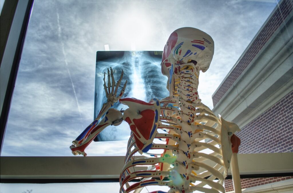
X-ray investigation is the original and most commonly used form of diagnostic imaging. Small amounts of radiation are passed through a selected part of the body to produce a diagnostic image. It is usually used to evaluate the chest and musculoskeletal system. This provides a view of an area where a patient is experiencing pain, monitors the progression of a disease, such as osteoporosis, and sees the effect of a treatment method, etc., all without making any incisions.
Imaging produced by X-rays shows different structures of the body in various shades of grey. In areas that don’t absorb X-rays well, the grey appears darkest. In dense areas that do absorb more of the X-rays, such as bones, the grey appears lighter. X-rays can be either still or moving images.
X-ray radiation is known as ionizing radiation. The dose of radiation involved is very low and is similar in strength to natural radiation that people are exposed to every day. Our radiographer always ensures the radiation dose is kept as low as possible and that the benefits of a patient having the X-ray outweigh any risks.
Our Services Include:
- X-RAY:
Skull PA/LAT
Mandible/Jaws
Sinuses
Mastoids
Soft Tissue Neck
Chest X-ray (PA)
Abdomen (AP)
Abdomen (Erect and Supine)
Pelvis
Extremities
Hand
Forearm (Radius & Ulna)
Arm (Humerus)
Foot
Leg (Tibia & Fibula)
Thigh (Femur)
Spine
Cervical
Thoracic
Lumbosacral
Joints
Wrist
Elbow
Shoulder
Ankle
Knee
Hip
Specials
HSG
IVU
Barium studies
Swallow
Enema
MCUG
RUG
2.ULTRASOUND:
Abdominal
Pelvic
Abdominopelvic
KUB
Female
Follicular Tracking
Transvaginal
Obstetric
First Trimester
2/3 Trimester
Biophysical
Gender Determination
Fetal well-being
Soft Tissue (Small Part)
Testicular
Thyroid
Breast
Prostate

